20 year old gentleman was GP-referred for ultrasound for a palpable lump in the left testis or
epididymis. The swelling was not painful, but of some discomfort.
Image Gallary:
Imaging findings:
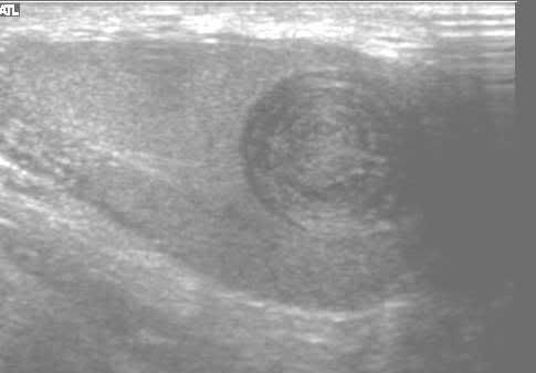
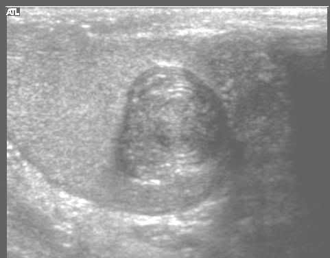
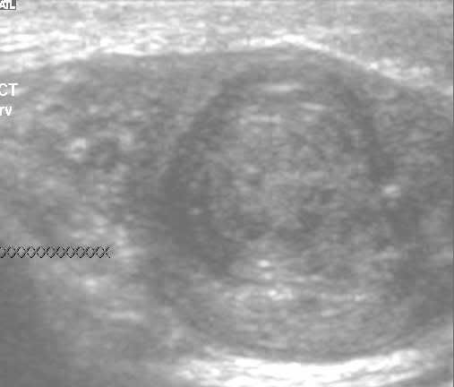
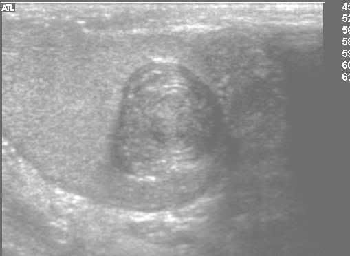
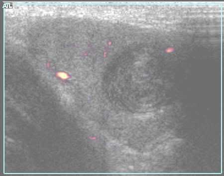
Ultrasound showed a solitary 2cm well defined round heterogenous echogenic mass in the inferior pole of the left testis. It showed rings of hypo- and hyper-echogenicities or 'onion-ring' appearance. On Doppler, there was no increased blood flow in or around the lesion. The mass was not related to the mediastinum of the testis. The epididymis was normal. The right testis and epididymis had normal appearances. There was no hydrocele or varicocele. The abdominal ultrasound showed no enlarged para-aortic lymphnodes. Based on the ultrasonic appearances, the presumptive diagnosis of the epidermoid cyst was made and further referral to the urologist was suggested (follow-up will be updated)
Discussion:
Epidermoid cyst is a rare benign lesion of the testis (1%–2% of testicular lesions). Commonly the patients present between 2nd and 4th decades. Most of the epidermoid cysts are single and unilateral. Multiple or bilateral cysts are associated with Gardner syndrome, Klinefelter syndrome and cryptorchid testes. They are filled with laminated cheesy material. The clinical management is controversial and recently, organ-preserving surgery has been favored over traditional orchidectomy.
Most patients present with painless mass, but a few complain of pain or discomfort. US may show an echogenic centre surrounded by a hypoechoic ring and hyperechogenic rim, causing 'bull’s-eye' or 'target' lesion; or alternating hypoechoic and hyperechoic concentric rings, causing 'onion skin appearance'. The lesions are not vascular.
MR shows low signal peripheral rim on both T1- and T2-weighted images and a circumferential high signal zone surrounding a low-signal central zone, or alternating concentric rings of low and high signal on T1- and T2-weighted images. On contrast, there is no enhancement.
The central echogenic center may represent keratin debris and the concentric layeers may represent lipid and water containing materials. The squamous cell capsule causes hyperechoic rim.
The 'onion ring' appearance is charecteristic for an epidermoid cyst, although not pathognomonic. Simple and tunica albuginea cysts are anechoic. Tumors, abscesses and chronic inflammatory processes may have capsule, but are likely to show hypervascularity. Neoplasms usually enhance on Gd-enhanced MR. Hemorrhage may have heterogenous appearance on ultrasound.
References:
1. Cho JH et al. Sonographic and MR Imaging Findings of Testicular Epidermoid Cysts. AJR 2002; 178:743-748
2. Loya AG et al. Epidermoid Cyst of the Testis: Radiologic-Pathologic Correlation. RadioGraphics 2004; 24: S243-S246.
3. Woodward PJ et al. From the Archives of the AFIP: Tumors and Tumorlike Lesions of the Testis: Radiologic-Pathologic Correlation. RadioGraphics 2002; 22: 189.
No comments:
Post a Comment