30 year old gentleman with known bowel carcinoma presented with obstrctive jaundice. On ultrasound there was marked intra and extrahepatic biliary dilatation, but no calculus or obvious mass was demonstrated. CT with pancreatic protocol was requested.
Image gallary:
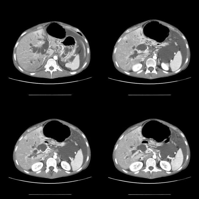
Arterial phase:
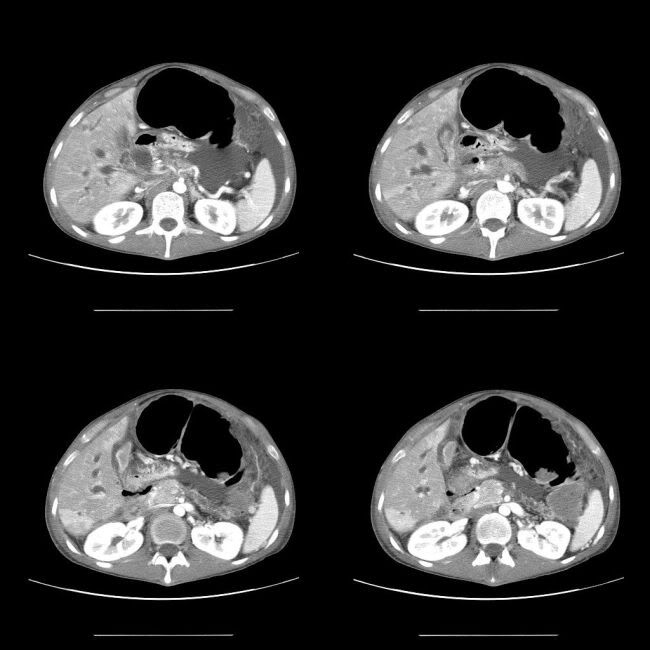
Venous phase:
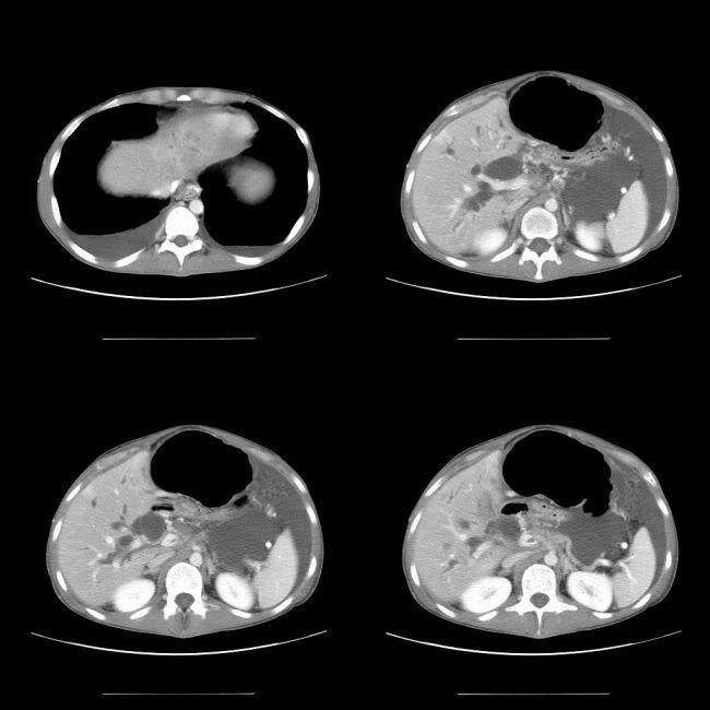
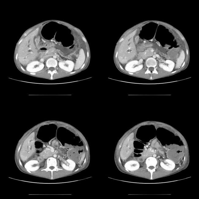
Obvious imaging findings:
- Marked intra and extra hepatic biliary dilatation
- Pancreatic duct dilatation in the body and tail region
- No obvious mass lesion in the head of the pancreas
Ancillary imaging findings, pointing the diagnosis:
- Encasement of the celiac axis
- Filling defect in the portal and superior mesenteric vein
- Oesophageal varices
- Ascites
Diagnosis:
Pancreatic mass causing marked biliary and pancreatic tree dilatation; encasing the celaic axis; thrombosis of the portal vein and SMV, leading to oesophageal varices and ascitis (portal hypertension)
Lesson:
1. pancreatic and biliary duct dilatation = pancreatic head/ampullary mass. If the mass is not obvious, look for ancillary signs, i.e.,
a. Celiac axis encasement
b. portal vein, SMV or/and splenic vein thrombosis (may be very subtle)
c. signs of portal hypertension
2. Always have a high level of suspision for pancreatic mass lesion.
No comments:
Post a Comment