Grading (American Association of Surgeons in Trauma)
1: parenchymal contusions, isolated subcapsular hematoma. 82% of renal injuries.
2: superficial cortical lacerations less than 1 cm deep, nonexpanding perirenal hematoma. 6%
3: lacerations greater than 1 cm deep without extension into collecting system or urinary extravasation. (3 & 4 - 7%)
4: Deep lacerations involving collecting system, traumatic thrombosis of segmental renal arterial branch, injury to main renal artery without devascularization
5: shattered kidney, devascularization - renal artery avulsion, main renal artery thrombosis (shearing injury to intima) (4 & 5 - 5%)
Radiological grading:
1: Minor injury: contusion, intrarenal or subcapsular hematoma, minor lacration with limited perirenal hematoma without extension into collecting system or medulla, subsegmental cortical infarct
2: Major: laceration extending from cortex to medulla/collecting system with/without extravasation, segmental infarct
3: Catastrophic: Multiple lacerations, pedicular injury
4: PUJ avulsion
Imaging findings:
On CT, contusions appear as a focal area of low attenuation with or without defined margins. Acute hematomas are of high attenuation. Small subcapsular hematomas are usually cresentic if small or lentiform if large. Laceration appears as a linear low attenuation. Superficials are less than 1cm and deep are more than 1cm deep. When renal capsule is lacerated, perinephric hemorrhage is usually occurs. Active hemorrhage or pseudoaneurysms are seen as intesne enhancing focal areas; active bleed tracks in the centre of surrounding hemorrhage; false aneurysm is round and focal. Active bleeding is a sign of decompensation (38% in one series) and are candidates for embolization. Urine leak is seen in delayed phase (pyelographic phase - 10 minutes later). Infarcts are wedge shaped low attanutaion traingular structures extending from medulla to cortex and do not enhance. Complete devascularization shows absent nephrogram or cortical rim nephrogram. Thrombosis or laceration of the renal vein is a rare and is type of renal pedicle injury.Venography is preffered as CT may not reliably detect venous laceration. Immediate surgical repair of venous injuries may be required to control bleeding. CT may reveal reveal intraluminal thrombus, nephromegaly, a diminished nephrogram, delayed nephrographic progression, and decreased excretion of contrast material into the collecting system, suggestive of acute venous hypertension.
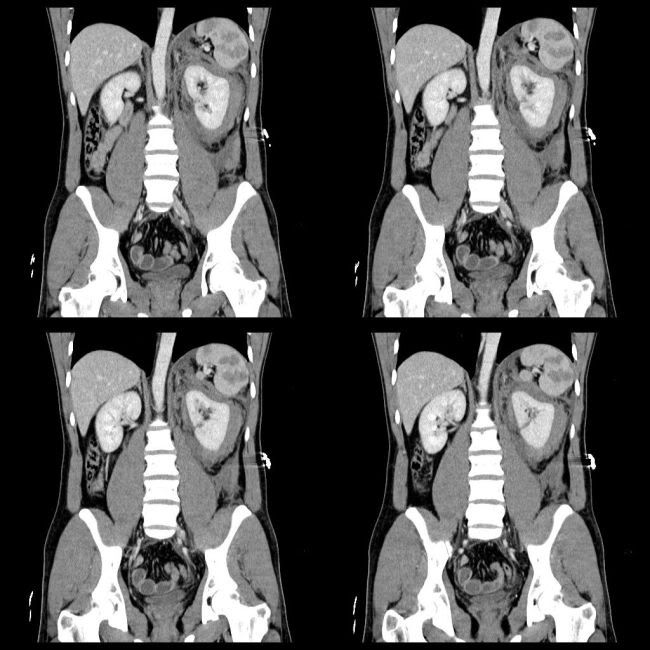
Image Gallary:
Management:
Grade 1 to 4 are managed conservatively. The only absolute surgical indication is life-threatening renal bleeding. Relative indications are presence of extensively devitalized tissue (>50% of parenchyma), uncontrolled urinary extravasation even by ureteral stent or nephrostomy, arterial thrombosis. Urine leak spontaneously resolve in up to 90%. Actively bleeding renovascular pedicle injuries (grade 5) need surgical exploration. Traumatic thrombosis or avulsion of renal artery needs repair within 4 hours with success of 14%–29%. If more than 4 hours with normal contralateral kidney they allow it to atrophy.Early complications occur within 4 weeks and include urinary extravasation and urinoma formation, delayed bleeding, infection of the urinoma, perinephric abscess, sepsis, arteriovenous fistula, pseudoaneurysm and hypertension. Late complications include hydronephrosis, hypertension, calculus formation and chronic pyelonephritis.
References:
1. Fanney DR et al.CT in the diagnosis of renal trauma. RadioGraphics 1990; 10: 29.S201-214
2. Kawashima A et al.Imaging of Renal Trauma: A Comprehensive Review. RadioGraphics 2001; 21: 57
No comments:
Post a Comment