75 year old lady with features of obstructive jaundice
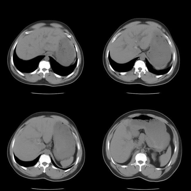
Unenhanced CT abdomen:
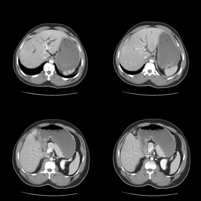
Enhanced CT abdomen:
Findings:
Marked intrahepatic biliary dilatation. A small area of calficiation and surrouding small area of low attenuation in the liver close to the caudate lobe. No extrahepatic biliary dilatation. No other abnormality. Unenhanced CT showed the findings better than the enhanced CT
ERCP:
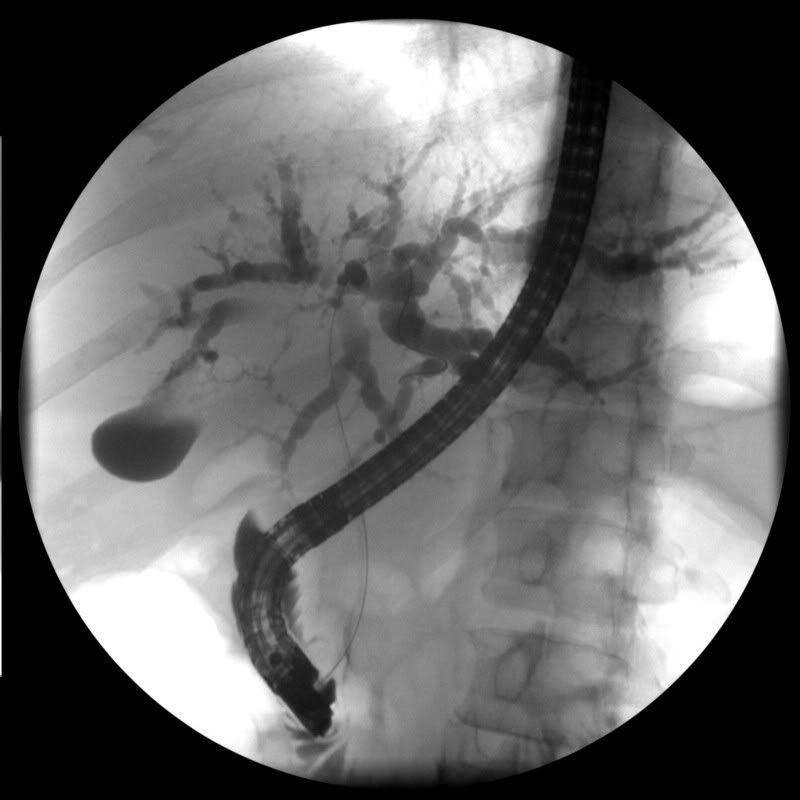
Findings:
Marked intrahepatic biliary dilataion. A focal area of irregular filling defect in the distal intrahepatic biliary segment
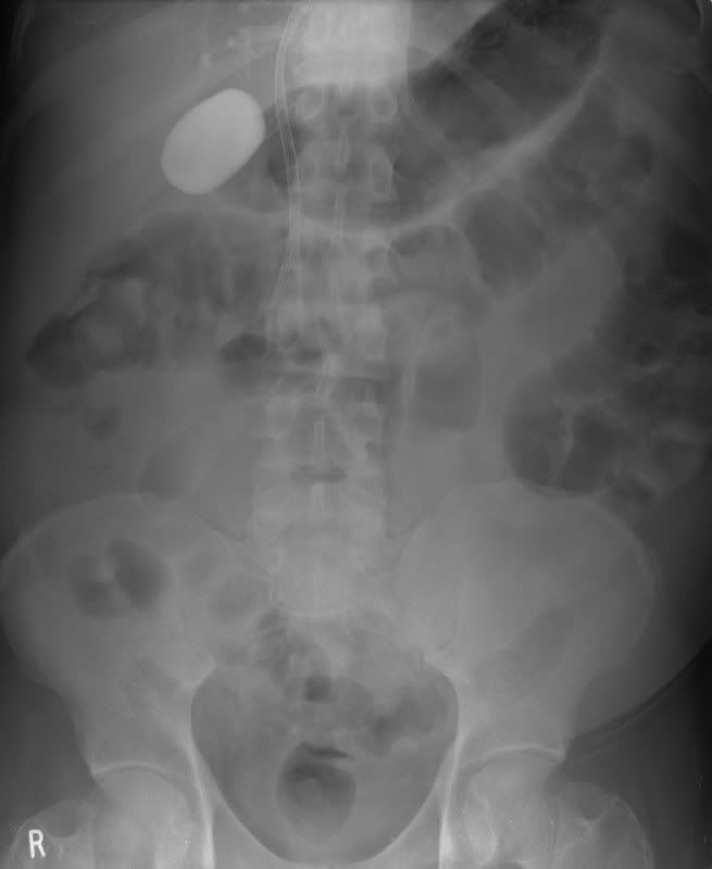
AXR: post percutaneous stenting
Diagnosis:
Intrahepatic cholangiocarcinoma
Discussion:
Calcification in intrahepatic cholangiocarcinoma is usally illdefined, mostly occurs in the periphery of the tumour and is seen in about 20% of patients. Calcifications may also be seen in in biliary cystadenocarcinomas. However, the most common cause of calcified hepatic lesions is inflammatory lesion like granulomatous diseases (TB). Hydatid cysts produce curvilinear or ring calcification. Large hemangiomas may show large central coarse calcffications. Hepatic adenoma may show solitary or multiple calcifications usually eccentric in location. Fibrolamellar carcinoma show calcifications in approximately 20% of cases . Calcfied hepatic metastases are most commonly due to mucin-producing neoplasms (colon carcinoma).
The likely causes for the calcifications in intrahepatic cholangiocarcinomas include central necrosis, mucinous type of cholangiocarcinoma. it is not known if the calcification can predict the prognosis of the disease.
Most of the cholangiocarcinomas are inoperable at the time of presentation and are treated with either ERCP or percutaneous stenting.
References:
1. Lee WJ et al. Radiologic Spectrum of Cholangiocarcinoma: Emphasis on Unusual Manifestations and Differential Diagnoses. RadioGraphics 2001; 21: 97
2. Stoupis C et al. The Rocky liver: radiologic-pathologic correlation of calcified hepatic masses.
RadioGraphics 1998; 18: 675
1 comment:
My battle with Emphysema started over 9 years ago which I finally got rid of with the help of Dr Itua herbal center treatment..I had the disease for over 9 + years..I'm in a good health now because Dr Itua herbal cure formula improve my condition drastically..the last time I went to the emergency PFT which is this year January I was told that my lung and breathing are working perfectly which was the help of this herbal medication..I don't have breathing problems anymore(Shortness of breath)..the Dr Itua herbal cure build up my lungs gradually after completing their prescription ,am able to cough it up no problem....I also met a lung specialist who told me that my lung is working perfectly so we don’t have to give it up because today i am here telling the world about my final victory with emphysema with the help of Dr Itua herbal cure and the help of their Natural herbal products and roots to cure and heal me completely from emphysema disease within the range of 5 weeks that I used the herbal medication. And if you have this kind of illness , there is no need to waste money on Corticosteroids or Zephyr Valve, or allowing doctors to waste their time on you instead why don’t you go get herbal products from Dr Itua Herbal Center use it and see for yourself And they also cures fibromyalgia,epilepsy,diabetes,cancer,pain killer,parkinson's,alzheimer’s disease,hiv/aids,herpes virus,hepatitis,pregnancy,and other diseases, contact him on drituaherbalcenter@gmail.com and www.drituaherbalcenter.com it very important you recommend this formula to anyone at there suffering from this illness people don’t know they exist .
Post a Comment