65 year old gentleman with progressive breathlessness
Image gallary:
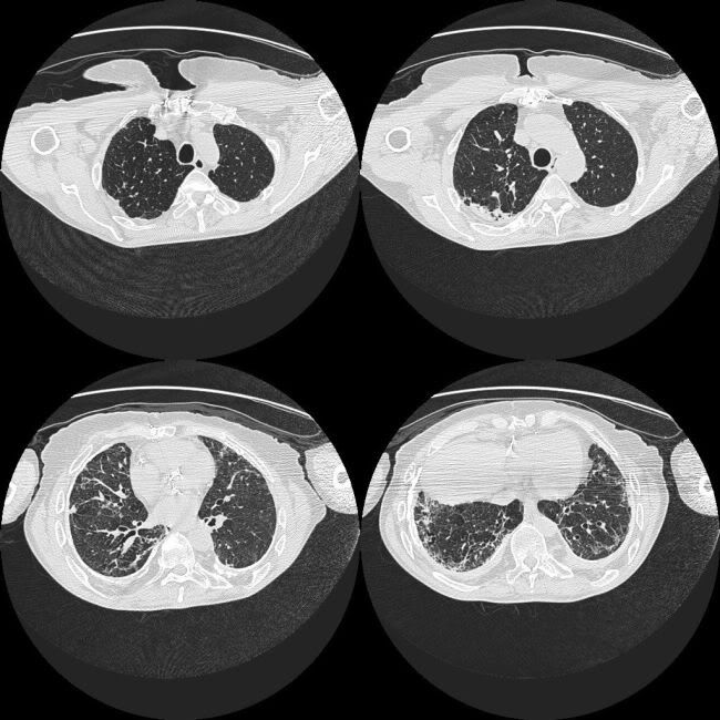
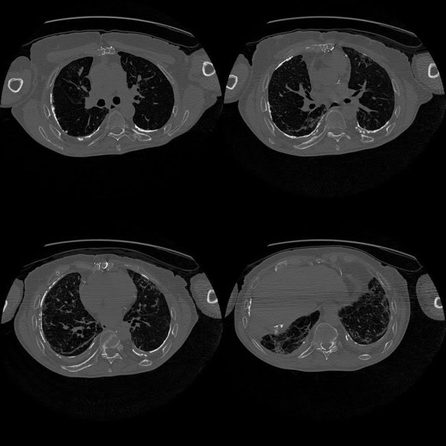
Findings:
HRCT lung windows: Bilateral basal interstitial lung disease - reticulations, fibrosis, architectural distortion, traction bronchiectasis
HRCT bone windows: Extensive calcified pleural plaques
Diagnosis: Diagnostic of asbestosis
Additional points to be noted/mentioned: Is there associated TB, pleural mass (mesothelioma)or lung mass (bronchogenic carcinoma)?
1 comment:
good case , just to add asbestos related calcification is usually bilateral while asbestos related fibrothorax is unilateral . however involves extensive involvement of pleurawhich may cover at least a fourth of the chest wall.
calcification associated with tuberculosis ( empyema)or resolved hemothorax of course would be on the side of previous lesion that is usually unilateral.
Post a Comment