58 year old gentleman presented with left ureteric colic. CT KUB was requested to exclude ureteric calculus. CT KUB was performed.
Images:
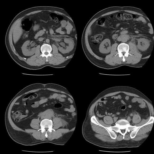
The case was reviewed by the reporting radiologist, before the patient was sent back. The reported radiologist asked for IV contrast enhanced study
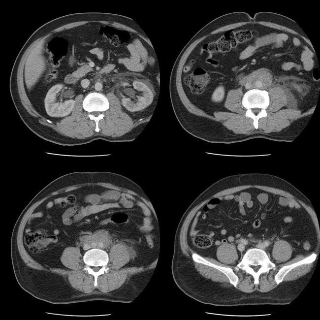
Findings:
Mild hydronephrosis of the left kidney with significant perinephric fat stranding. The right renal pelvis and both ureters are also slightly prominant upto the level of the retroperitoneal mass lesion encasing the aorta, IVC and both ureters. No similar masses were found the in the abdomen or pelvis.
Diagnosis:
Retroperitoneal fibrosis
Discussion:
Retroperitoneal fibrosis (RPF) usually presents with a dull aching non-colicky pain in the flank, back, scrotum or lower abdomen. Other symptoms may include fever, ankle edema and DVT. Uncommon presentations include weight loss, nausea, vomiting, anorexia and malaise.
Rarely RPF may present with ureteric colic (as in the present case), Raynaud phenomenon, hematuria, claudication or urinary frequency.
60-70% are idopathic, but can be associated with malignancy, inflammatory processes (Crohn disease, ulcerative colitis, sclerosing cholangitis), trauma, radiation, drugs (methysergide, beta blockers, metyldopa). Steroids are used in the management of the disease.
No comments:
Post a Comment